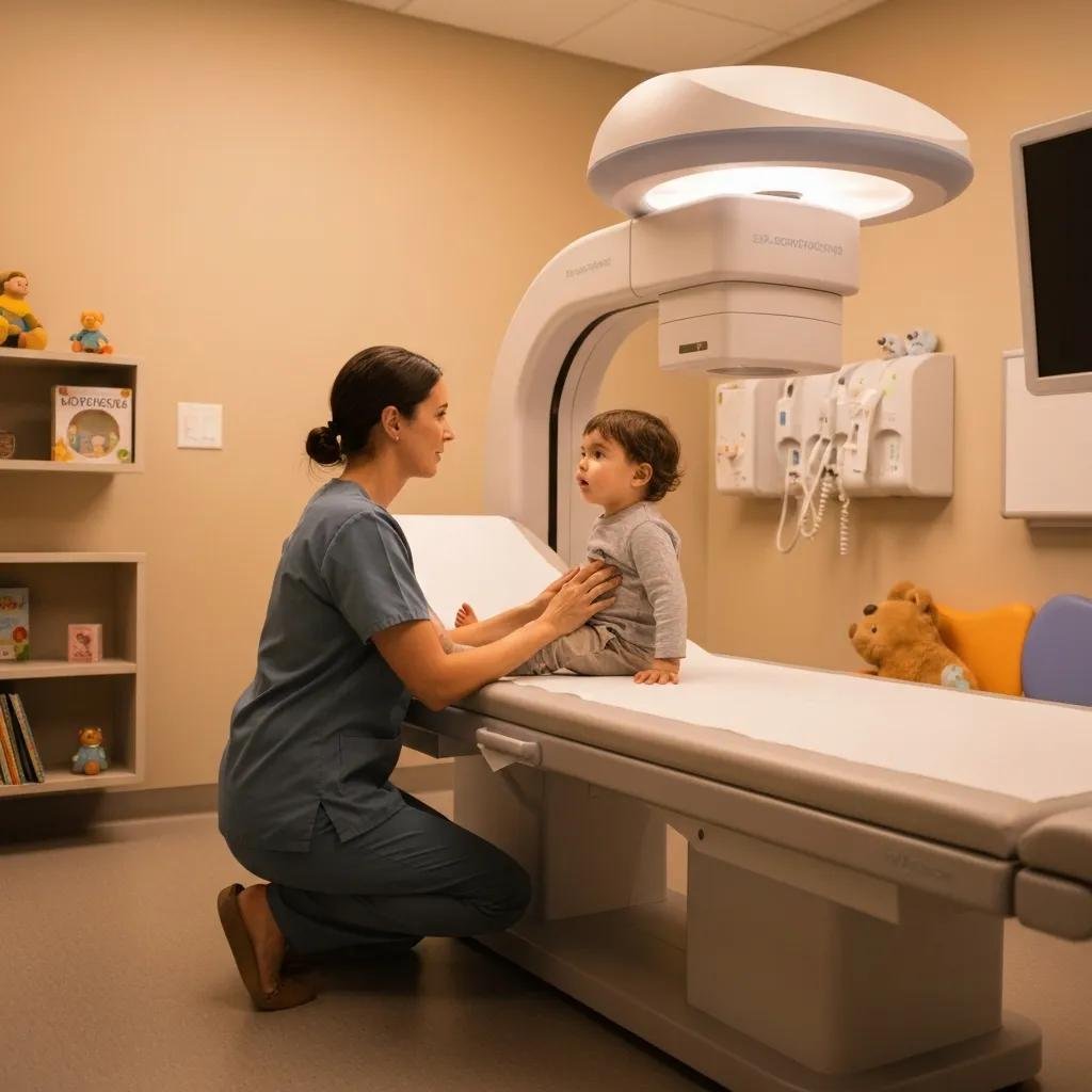
Bone age assessment measures how far a child’s skeleton has developed compared to typical growth patterns. Physicians use it to evaluate growth disorders, hormonal conditions, and timing of puberty. The process relies on pediatric imaging.
Common Methods
Several established techniques guide bone age assessment in pediatric imaging. Each approaches skeletal maturity from a slightly different angle. The Greulich and Pyle method compares a single hand-and-wrist radiograph against a reference atlas. The reader matches the patient’s film to the closest standard image, then assigns a corresponding bone age. This method remains widely used because it is fast and straightforward.
The Tanner-Whitehouse method takes a more detailed approach. It scores individual bones based on their stage of maturity, then combines those scores into an overall rating. This method may take longer to complete, but it can reduce variation between readers.
Newer approaches use automated software to analyze radiographs. These systems measure bone features and generate an estimate based on large reference datasets. Automated tools can support consistency, though a trained radiologist still reviews the final result.
Standard Age Assessment
Standard assessment begins with a single radiograph of the left hand and wrist. The left side is used by convention, which keeps results comparable across patients and studies. The radiologist examines specific structures during interpretation:
- The growth plates at the ends of the long bones
- The carpal bones in the wrist
- The shape and fusion of the bones in the fingers and thumb
These features change in a predictable sequence as a child matures. The reader compares them against established reference standards to estimate skeletal maturity. The result is reported as a bone age, which is then compared to the child’s chronological age.
A meaningful gap between bone age and chronological age can point to a growth or hormonal condition. Children whose bone age is well ahead of chronological age may be experiencing early maturation. Children whose bone age lags behind could have delayed development. These findings guide further evaluation rather than provide a diagnosis on their own.
Some Considerations
Skeletal maturity can vary across ethnic groups, by sex, and over generations. The patient’s age range matters as well. Different bones provide the clearest signals at different stages of growth. In very young children, the carpal bones offer useful information. In older children and adolescents, the growth plates and their fusion become more telling.
Finally, bone age is one part of a broader clinical picture. Results carry the most value when combined with height, weight, medical history, and other findings. A single measurement should not stand alone in clinical decisions.
Schedule Pediatric Imaging
If your child has been referred for a bone age study, accurate imaging starts with the right preparation. Ask your provider which technique the imaging center uses and whether a trained pediatric radiologist will interpret the results. Contact your imaging center to schedule the study and bring any prior records that can help with comparison. A well-documented assessment gives your child’s care team some of the information needed to plan the next steps.
About the author : ahmadrazaguruu@gmail.com